 |
Viсtoriya V. Zherdeva Ph.D. (Biology) Head of Laboratory INBI, build. 2, room 118 |
General
Key words:
molecular imaging, time-resolved fluorescence, magnetic resonance imaging, proteinases, fluorescent proteins, genetically encoded sensors, inflammation, carcinogenesis
History of the lab foundation:
The Molecular Imaging Laboratory (MIL) and its research team were established in March of 2018 in accordance with the Decree No. 220 of the Government of the Russian Federation from April 9, 2010. The laboratory is supported by the Grant No. 14.W03.31.0023 from the Ministry of Education and Science of Russian Federation entitled “Visualization and engineering of eukaryotic genomes”(“Megagrant”). This Grant was awarded under the auspices of the Institutional development of the research sector within the Russian Federation Government Program “Development of Science and Technology” for the period of 2013-2020.
![]() PROJECT INFORMATION “Visualization and engineering of eukaryotic genomes”
PROJECT INFORMATION “Visualization and engineering of eukaryotic genomes”
Research
RESEARCH
Laboratory of Molecular Imaging was established with the purpose of solving a number of important scientific problems related to the field of medical biotechnology and nanotechnology. MIL was created on the basis of a scientific team from the Federal State Research Center “Fundamental Foundations of Biotechnology “of the Russian Academy of Sciences. The researchers participating in the project have prominent international standings in their respective areas, and are experts in their fields, i.e. non-invasive imaging in living systems, biophysics, biochemistry and engineering of fluorescent proteins, as well as radiology.
We created an interdisciplinary research team in order to obtain breakthrough results within the framework of several interrelated directions of research at the junction of molecular biology, biophysics and medicine. The main goal of the laboratory is to achieve breakthroughs in the field of mapping eukaryotic genome imaging using multimodal / tomographic and multidisciplinary approaches. Scientific breakthroughs are expected as a result of the convergence of efforts to unite researchers who are currently working in parallel areas with a high level of significance and innovation, thereby strengthening the overall scientific potential. The laboratory is actively engaged in research along the following directions:
- development of bioengineering approaches for creating fluorescent sensor molecules based on mutants derived from the Cas family of endonucleases;
- application of the achievements of the laboratory of the leading scientist in the field of nanotechnologies for the delivery of recombinant molecules and viral constructs for the purpose of regulated expression of CRISPR / Cas9 based sensors in cells and tissues of experimental animals;
- the use of picosecond laser technology to study the fluorescence lifetimes of recombinant sensor molecules in the context of targeting genomic DNA;
- the creation of a hybrid imaging technology that combines magnetic resonance imaging and fluorescence visualization of lifetimes in time domain.
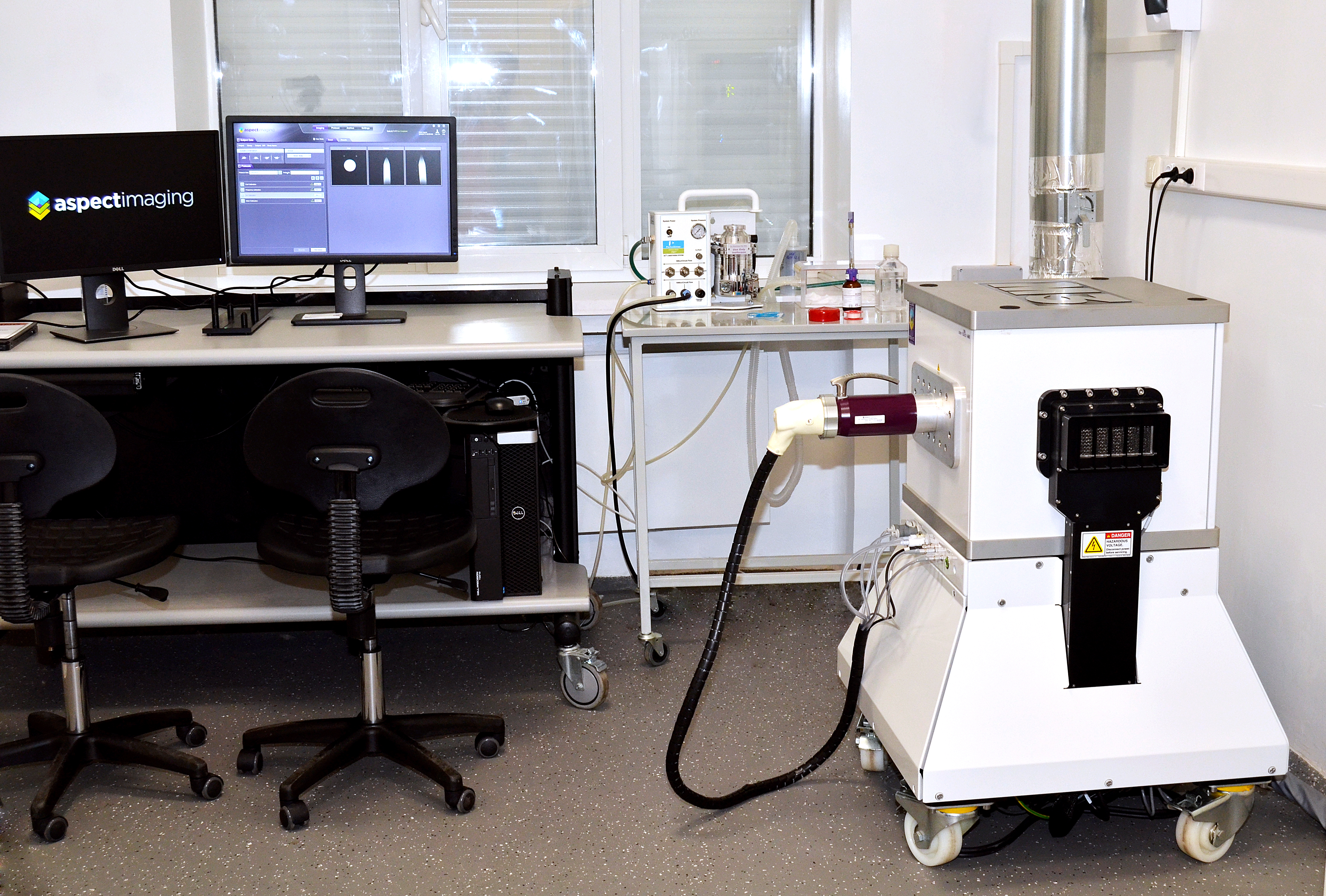
In 2019 MIL received and installed the latest version of M3 ™ preclinical magnetic resonance imaging (MRI) unit (Aspect Imaging, Shoham, Israel). The device is designed to visualize the details of the anatomy and physiology of experimental animals. The M3 ™ MRI is based on a compact, highly efficient permanent magnet with a magnetic field strength of 1 Tesla, which does not require cryogenic support. This MRI setup is one of about 100 preclinical MRI systems installed around the world. This MRI allows to obtain anatomical and functional tomographic images with high spatial resolution (i.e. voxel volume), reaching fractions of a cubic millimeter (microliter). The system includes a system for anesthesia and immobilization of the animal, a physiological monitoring system, as well as all the necessary components for collecting images and image analysis, including computer software for imaging obtained in multi-channel, multi-modal operating modes in combination with positron emission and optical tomography.
Facilities and equipment
FACILITIES AND EQUIPMENT
 At the end of May 2019, as part of the implementation of the project No. 14.W03.31.0023 of the Ministry of Science and Higher Education of the Russian Federation, the latest M3 ™ device for preclinical magnetically was installed and commissioned in the laboratory of the INBI Molecular Imaging Laboratory (bldg. 2, room 120) – resonance imaging (MRI). The device was manufactured by Aspect Imaging (Shoham, Israel) and is designed to visualize the details of the anatomy and physiology of experimental animals (rodents weighing up to 30 g).
The M3 ™ MRI is based on a compact, highly efficient permanent magnet with a magnetic field strength of 1 Tesla, which does not require cryogenic support. This MRI setup is still the only system in Russia, and one of about 100 preclinical MRI systems installed in leading research and R&D centers in the world.
The tomographic images obtained using this MRI device are based on differences in relaxation times and concentration of protons of molecules, in a large number of living organisms present in the tissues. Image data can be obtained with high volume resolution (i.e. voxel volume), reaching fractions of a cubic millimeter (microliter).
The system includes a system for anesthesia and immobilization of an animal, a physiological monitoring system, as well as all the necessary components for obtaining, recording, collecting data and analyzing them, including computer software for recording images obtained in multi-channel, multi-modal operating modes, including combination with positron emission and optical tomography.
This system meets the high needs of the laboratory in combining approaches for obtaining MRI images and fluorescence images on the same experimental animal to solve the problems of visualization and localization of various processes in a living organism in dynamics, normal and pathology.
Publications
PUBLICATIONS
Selected publications:
- Bogdanov A.A., Tuchin V.V., Meerovich I.G., Kazachkina N.I., Zherdeva V.V., Solovyev I.D., Tuchina D.K., Savitsky A.P. Towards registration of optical and MR signal changes in subcutaneous tumor volume in vivo after optical skin clearing. В сборнике: Progress in Biomedical Optics and Imaging – Proceedings of SPIE. Сер. “Dynamics and Fluctuations in Biomedical Photonics XVII” 2020. С. 112390N. (SJR IF 0,27)
- Bogdanov AA Jr, Meerovich I, Kazachkina N, Zherdeva V, Solovyev I, Tuchina DK, Savitsky AP, Tuchin VV. Optical clearing effects in subcutaneous red-fluorescent tumors monitored by fluorescence and magnetic resonance imaging in vivo (Conference Presentation). SPIE Proceedings Volume 11363, Tissue Optics and Photonics; 113630S (2020) https://doi.org/10.1117/12. 2560186 SPIE Photonics Europe, 2020, Online Only, France April 6-8 (SJR IF 0,27)
- Daria K. Tuchina, Irina G. Meerovich, Olga A. Sindeeva, Victoria V. Zherdeva, Alexander P. Savitsky, Alexei A. Bogdanov, Jr Valery V. Tuchin. Magnetic resonance contrast agents in optical clearing: Prospects for multimodal tissue imaging. J.Biophotonics. 2020 https://doi.org/10.1002/jbio. 201960249 (2021-SJR Q1 IF 0,8)
- A.A. Bogdanov Jr., N.I. Kazachkina, V.V. Zherdeva, I.G. Meerovich, D.K. Tuchina, I.D. Solov’ev, A.P. Savitsky, V.V. Tuchin Optical clearing and multimodality fluorescence and magnetic resonance imaging in cancer models Proceedings of SPIE, 2021, Vol. 11641, P.1-8 DOI: 10.1117/12.2586113 (SJR IF 0,18)
- Kazachkina NI, Zherdeva VV, Saydasheva AN, Meerovich IG, Tuchin VV , Savitsky AP, Bogdanov AA Jr.Topical Gadobutrol Application Causes Fluorescence Intensity Change in RFP-expressing Tumor-Bearing Mice. J. Biomedical Photonics & Eng 2021 7(2) 020301-1. (2021 – SJR Q2 IF 0.39)
- Tuchina DK, Meerovich IG, Sindeeva OA, Zherdeva VV, Kazachkina NI, Solov’ev ID, Savitsky AP, Bogdanov AA Jr., Tuchin VV. Prospects for multimodal visualisation of biological tissues using fluorescence imaging. Quantum Electron 2021 51(2) 104. ( 2021- SJR Q2 IF 0.4)
- Bogdanov AA Jr, Kazachkina NI, Zherdeva VV, Meerovich IG, Tuchina DK, Solovyev ID, Savitsky AP, Tuchin V.V. Magnetic resonance imaging study of diamagnetic and paramagnetic agents for optical clearing of tumor-specific fluorescent signal in vivo. Chapter 15 In: Tissue optical clearing: new prospects in optical imaging (Valery Tuchin, Dan Zhu, Elina A. Genina(Eds.) CRC Press, Boca Raton, FL 2022.
- Zherdeva V, Kazachkina NI, Shcheslavskiy V, Savitsky AP. Long-term fluorescence lifetime imaging of a genetically encoded sensor for caspase-3 activity in mouse tumor xenografts. Journal of Biomedical Optics. 2018; 23(3): 035002. DOI: 10.1117/1.JBO.23.3.035002.
- Sdobnov AY, Darvin ME, Schleusener J, Lademann J, Tuchin VV. Hydrogen bound water profiles in the skin influenced by optical clearing molecular agents-Quantitative analysis using confocal Raman microscopy. J Biophotonics. 2019;12(5):e201800283. doi: 10.1002/jbio.201800283. Epub 2019 Jan 4. (WoS, Q1, IF 3.768, SJR 1.03)
- Sdobnov AY, Lademann J, Darvin ME, Tuchin, VV. Sensors for Proteolytic Activity Visualization and Their Application in Animal Models of Human Diseases, Biochemistry (Moscow), 2019; 84, S144-S158. WoS, Q4, I F 1.724, SJR 0.16)
- Bogdanov AA Jr, Solovyev ID, Savitsky AP. Sensors for Proteolytic Activity Visualization and Their Application in Animal Models of Human Diseases. Biochemistry (Moscow), 2019; 84, S1-S18. (WoS, Q4, I F 1.724, SJR 0.16)
- Munkhbat O, Canakci M, Zheng S, Hu W, Osborne B, Bogdanov AA, Thayumanavan S. 19F MRI of polymer nanogels aided by improved segmental mobility of embedded fluorine moieties. Biomacromolecules, 2019 DOI: 10.1021/acs.biomac.8b01383.
- Bogdanov A, Metelev V, Taghian T, Solovyev I, Kumar ATN, Zhang S, Savitsky AP. Near-infrared oligonucleotide duplex sensors for imaging rapidly activated transcription factors in vitro and in situ Paper No. 10877-17 SPIE Digital Library 2019.
- Leporati A, Gupta S, Bolotin E, Castillo G, Alfaro J, Gottikh MB, Bogdanov, Jr. AA. Antiretroviral hydrophobic core graft-copolymer nanoparticles: the effectiveness against mutant HIV-1 strains and in vivo distribution after topical application. Pharm Res, 2019; 36: 73. https://doi.org/10.1007/s11095-019-2604-9
- Rodríguez-Rodríguez A, Shuvaev S, Rotile N, Jones CM, Probst CK, Dos Santos Ferreira D, Graham-O′Regan K, Boros E, Knipe RS, Griffith JW, Tager AM, Bogdanov A Jr, Caravan P. Peroxidase Sensitive Amplifiable Probe for Molecular Magnetic Resonance Imaging of Pulmonary Inflammation. ACS Sensors 2019/08/09 DOI:10.1021/acssensors.9b01010
- Xie Q, Zeng N, Huang Y, Tuchin VV, Ma H. Study on the tissue clearing process using different agents by Mueller matrix microscope. Biomed Opt Express. 2019 Jul 1; 10(7): 3269–3280. doi: 10.1364/BOE.10.003269
- Taghian, T, Metelev, VG, Zhang, S, Bogdanov, AA Jr. Imaging NF-κB activity in a murine model of early stage diabetes. The FASEB Journal. 2020;34:1198–1210.
- Metelev VG, Bogdanov AA Jr. Synthesis and applications of theranostic oligonucleotides carrying multiple fluorine atoms. Theranostics 2020; 10(3):1391-1414. doi:10.7150/thno.37936 IF 8.0